Conventional histology
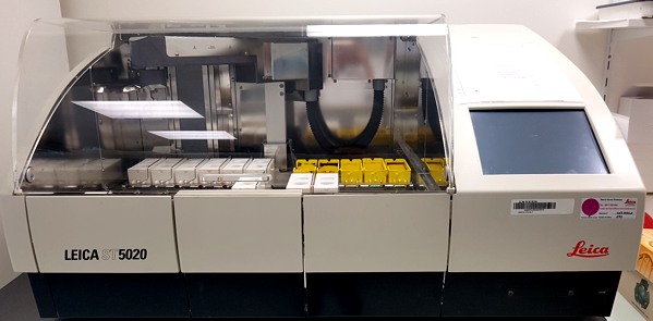
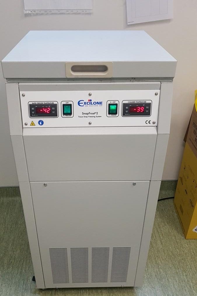
Tissue preservation (freezing, fixation), paraffin inclusion, slicing, histological staining

Methodological advice as well as assistance in the reading and interpretation of histomorphological and immunohistochemical results.
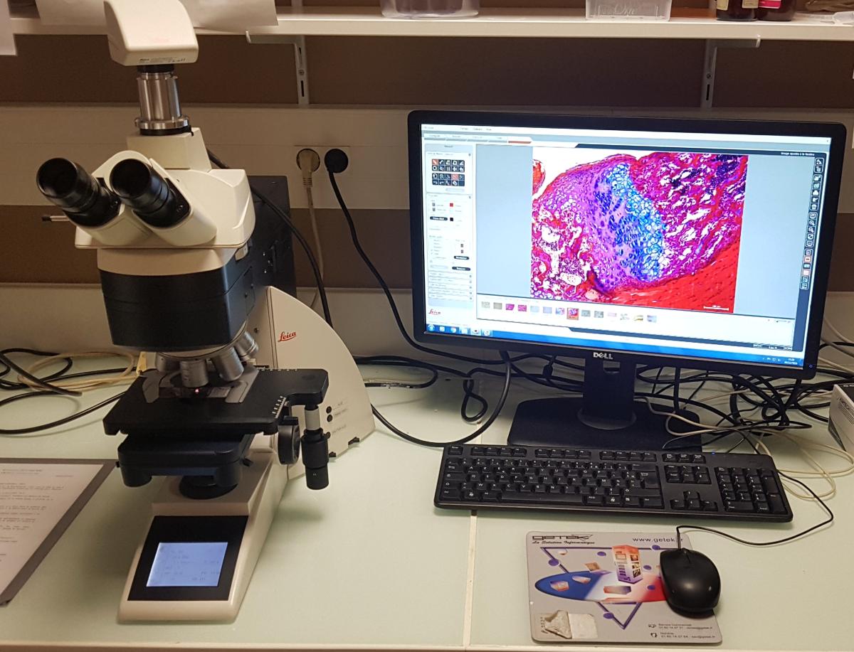
Equipment is made freely available after training in sample preparation, histological cutting and staining, immunolabelling, the use of microscopes, and slide digitization and analysis.
Open to academics as well as private companies, the department is involved in scientific collaborations, R&D, service delivery, consulting, and training.
Tissue preservation (freezing, fixation), paraffin inclusion, slicing, histological staining
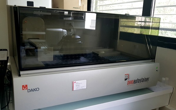
Immunohistochemistry/immunofluorescence of tissue sections (freezing, paraffin).
Fine-tuning the conditions for antibody use.
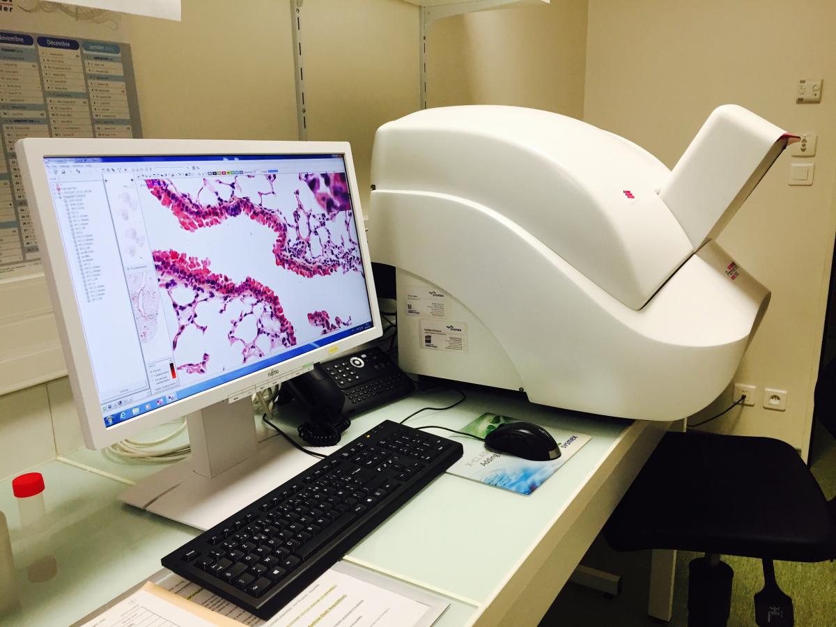
Digitization and quantification of histological slides with a Pannoramic 250 (3DHistech) slide scanner and associated software.
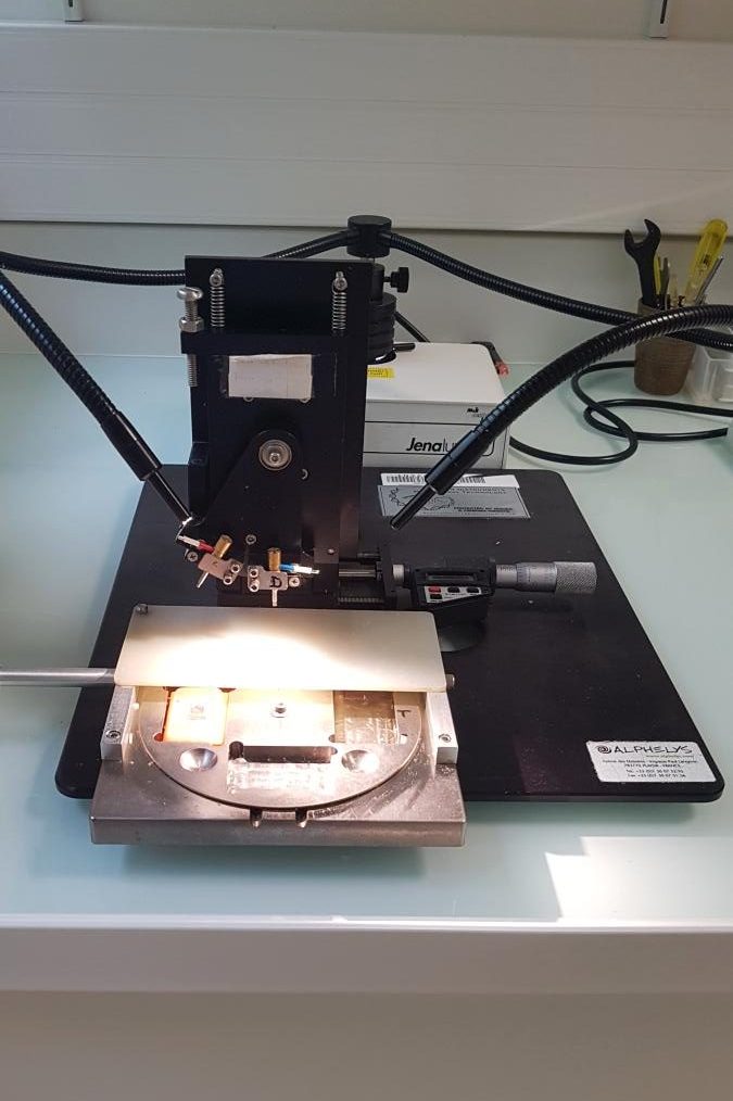
[TMA] using a Beecher Instruments tissue microarrayer
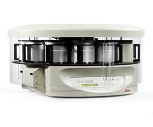
Dehydration
Slide scanner with quantification software
Tissue freezing
Making of tissue microarrays
Cryosectioning
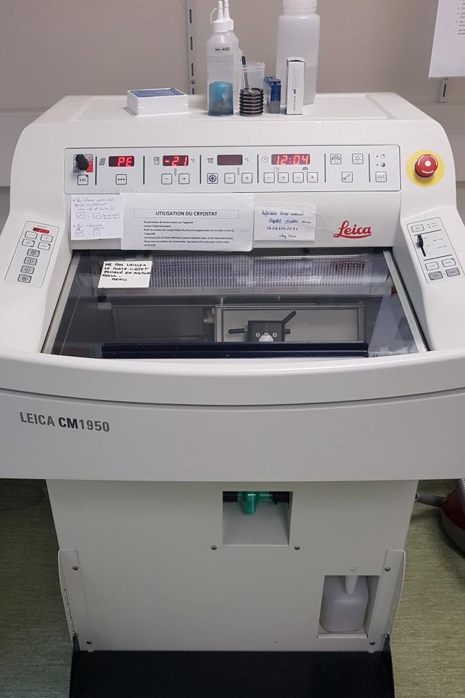
Paraffin block slicing
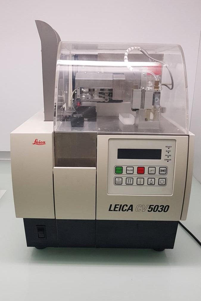
Paraffin block slicing
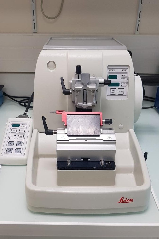
Automated immunohistochemistry system
